Authors: Shikha Malhotra, MBBS; Bryan Rea, MD; Nidhi Aggarwal, MD
Clinical History
A 61-year-old man presented with multiple chest wall masses. Past medical history was significant for type II diabetes, hypertension, hyperlipidemia, nephrolithiasis, basal cell carcinoma (status post excision), and bilateral testicular masses (status post recent bilateral orchiectomy at an outside institution 2-3 months ago).
Biopsy of one of the right chest/shoulder nodules was performed. On gross examination, the mass was rubbery to firm and measured 2.7 x 1.8 x 1.0 cm with the attached adipose tissue. Cut surface was fleshy, tan with small hemorrhagic areas. Histological sections demonstrated sheets of a monotonous population of cells infiltrating into adjacent adipose tissue. The neoplastic cells showed irregular nuclear contours, single to multiple prominent nucleoli and multiple apoptotic bodies. Paraffin section immunohistochemical stains performed at UPMC revealed the lesional cells to be positive for IRF8, CD68 (weak), CD33 and NPM1, and negative for CD34, CD117, MPO (myeloperoxidase), glycophorin, E-cadherin, CD61, CD19, CD20, PAX5, CD3, and pankeratin. Ki67 was high at 70%. P53 showed scattered positive cells. Flow cytometry studies revealed an expanded myelomonocytic population which was positive for CD123 (dim), CD33, HLA-DR, CD56, CD64, CD36, CD13 (dim, variable) and negative for CD34, CD117, CD16, CD11b, CD14 and TdT. The specimen yielded only three metaphases for chromosome analysis which showed trisomy 8 and an abnormal chromosome 7 with additional material attached to the short arm. A NGS Panel for mutations revealed NPM1 p.W288fs (c.863_864insTAAA, VAF 44.8%), FLT3 p.I836del (VAF 42.9%) and PTPN11 p.E76A (VAF 50.1%) mutations.


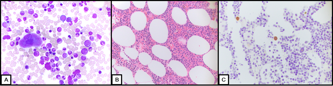
Additionally, peripheral blood and bone marrow evaluation was also performed. Complete blood count and peripheral blood flow cytometry studies were unremarkable at presentation. Bone Marrow was normocellular for age with trilineage hematopoiesis and no overt increase in blasts. Conventional cytogenetic studies, microarray analysis, fluorescence in-situ hybridization (FISH), and myeloid NGS (a 50-gene targeted DNA sequencing panel) performed on the bone marrow, were negative for an abnormal clonal population. However, in view of the biopsy results from the right chest/shoulder mass, additional immunohistochemical studies were performed which identified rare cells (<1% of total cellularity) with NPM1 positivity.